
Please visit Melbourne Gallbladder Centre
Gallbladder Disease
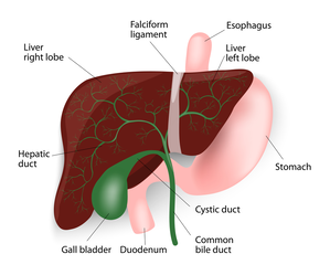
The gall bladder is a small pear-shaped organ attached to the side of the bile duct by a small secondary duct, the cystic duct. The bile duct is a tube that carries bile from the liver to the small intestine. When the patient is fasting, the lower end of the bile duct closes and bile back-flows into the gall bladder. There it is concentrated by the gall bladder, absorbing the water in the bile. When a patient eats a fatty meal, the gall bladder squeezes out the bile to help absorb the fats. If stones are present, they can cause the gall bladder to go into spasm and causes severe pain as well as nausea, vomiting, bloating, or fever. If a gallstone travels down the bile duct, you could have liver dysfunction, bile duct infection or inflammation in the pancreas, called pancreatitis. The only way to prevent this and the other problems that can occur with gall stones is to remove the gall bladder. Since the gall bladder is only one of the mechanisms of fat digestion, its removal does not cause any major interference with the patient’s digestive process. In many cases of patients with stones, the gall bladder is not functioning and so digestion of fats is not affected by its removal.
Gallstones Vs. Gallbladder Cancer
Although gallstones are a common problem, gallbladder cancer is a rare form of cancer except in certain parts of the world and in some ethnic groups. Underlying genetic or dietary factors may be the reason for this.
Gallstones are a risk factor for gallbladder cancer, though the mechanism is unclear. Usually patients with large stones of long duration are at increased risk for cancer. More than the stones themselves, incomplete emptying of the gallbladder and the chronic inflammation that goes with it may have a role to play in initiating the cancer.
Calcium deposits in the wall of the gallbladder (a consequence of chronic inflammation), described as Porcelain gallbladder, increase the risk of gall bladder cancer. The picture on this page shows a CT scan, with a porcelain gall bladder clearly visible (you can click on the picture to enlarge it).
Chronic Salmonella infection of the gallbladder, which predisposes to gallstone formation, can predispose to gallbladder cancer.
Patients with choledochal cysts (a focal or diffuse bulge in the bile duct) or abnormalities at the point where the pancreatic and bile ducts join and enter the bowel are also known to be at increased risk for gall bladder or bile duct cancer.
Sometimes patients are found to have polyps (little growths) on the lining of the gallbladder. Polyps are generally benign, but patients with a single, large (> 1cm) polyp are more likely to develop cancer within their polyp. See below for more information about gall bladder polyps.
Gallbladder Polyps
Polyps are growths on the inner lining of the gallbladder wall. Gallbladder polyps are often discovered incidentally on scans, and not all of them warrant removal. Polyps that are single, sessile (do not have a stalk), and are 1 cm or more in diameter should be deemed as suspicious i.e. they may develop into cancerous (malignant) growths. Also, polyps that have developed in older patients (over the age of fifty years), developed in association with stones or are associated with symptoms, are at a higher risk of being malignant or subsequently turning malignant. Laparoscopic cholecystectomy (keyhole surgery to remove the gall bladder) is the treatment of choice in these patients, unless the suspicion of malignancy is high, in which case open exploration is preferable, with preparation for extended resection if necessary.
For polyps that are deemed low-risk, Mr Karametos's practice is to recommend follow-up at first with regular ultrasound scans every 6-12 months, which can stop after 2 years if they remain unchanged. Mr Steven Karametos also offers minimally invasive laparoscopic surgical options for common bile duct exploration or stone extraction.
For polyps that are deemed low-risk, Mr Karametos's practice is to recommend follow-up at first with regular ultrasound scans every 6-12 months, which can stop after 2 years if they remain unchanged. Mr Steven Karametos also offers minimally invasive laparoscopic surgical options for common bile duct exploration or stone extraction.
Other Problems With Similar Gallstone Symptoms
The symptoms of gallstones can be similar to those of heart disease, appendicitis, bowel obstruction, peptic ulcer, hiatus hernia, pancreatitis, hepatitis and occasionally biliary cancer. It is therefore very important that the correct diagnosis is made. Other tests may be required, including an Gastroscopy to look inside the oesophagus and the stomach.
Read More About...